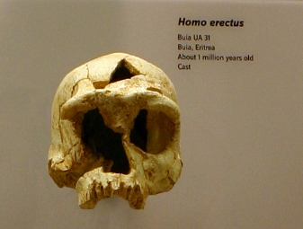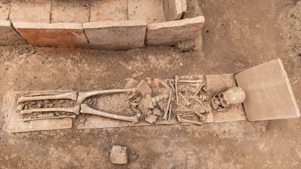
Most people picture a “runner’s skeleton” as something modern – carbon-fiber shoes, marathon medals, maybe a stress fracture from a treadmill. But bioarchaeologists have spent decades proving that a lifetime of walking rewires the human skeleton in ways that are still visible thousands of years after death. Bone isn’t static. It bends, thickens, flattens, and scars itself around the exact stresses a body endured, and it keeps that record long after the person carrying it is gone.
Some skeletons genuinely seem to remember every mile they ever walked. Forensic anthropologists can look at a femur, a hip socket, or even a single healed foot fracture and reconstruct a walking life most of us would find unimaginable today. Here are the thirteen tell-tale signs scientists actually rely on – and by the end, you’ll never look at a museum skeleton case the same way again.
#1 – The Femur Shaft Is Unusually Thick and Rounded
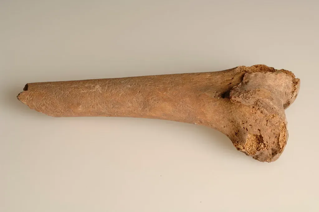
The thigh bone is the loudest storyteller in the human body when it comes to mobility. Researchers use a technique called cross-sectional geometry – essentially slicing a femur virtually, using CT scans, to measure how much bone material surrounds the marrow cavity and how that material is spread out. Bone is constantly working to minimize the stress running through it, and nowhere is that truer than in the legs, which is exactly why the lower limb skeleton is so reactive to shifts in daily activity.
A skeleton that walked constantly across long distances typically shows a femur that isn’t just long, but structurally reinforced for bending resistance. This isn’t the same as simply having “big bones.” It’s targeted reinforcement, concentrated exactly where the mechanical strain from repetitive striding would have been highest.
The bone literally builds armor around the load path of a walking gait. Forensic teams measure something called the “polar second moment of area,” a way of scoring how well a bone resists twisting and bending forces from repeated impact. High-mobility individuals consistently score higher here than sedentary counterparts, even after body size is factored out entirely.
#2 – The Femur Is Flattened Front-to-Back (Platymeria)
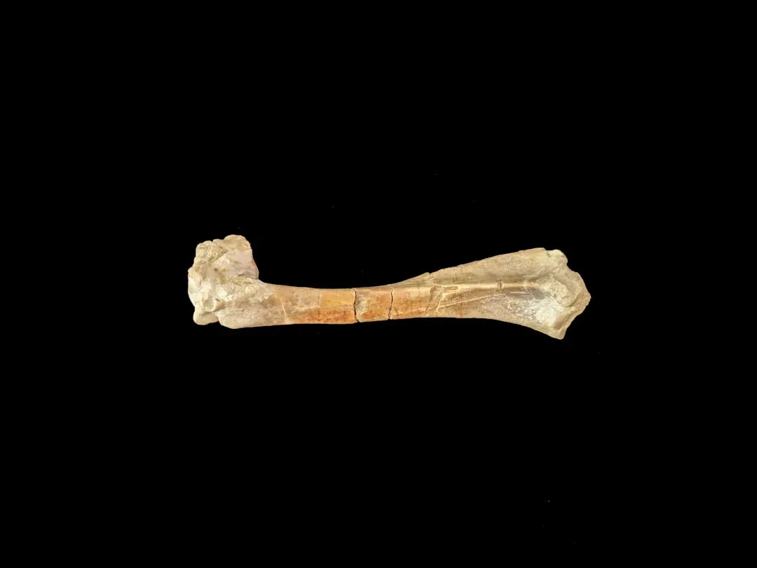
Here’s something most people have never heard of: long-distance walkers often develop a femur that’s flattened in one specific direction, called platymeria. Instead of a round, pillar-like shaft, the bone becomes oval in cross-section – flattened front-to-back rather than side-to-side.
This isn’t random. It reflects the exact bending forces walking places on the thigh bone during the stance and swing phases of gait. Anthropologists studying rugged, mountainous populations found the femur adapts far more aggressively than the same bone in flat-terrain populations. The steeper and rougher the terrain a person regularly crossed, the more pronounced this flattening becomes.
Classic comparative research on mountainous populations found that the added femoral strength in these groups lines up almost perfectly with the predicted mechanical cost of traveling over rough terrain – while the arm bones barely react at all, since the humerus does very little locomotor work during normal walking. That detail matters enormously for identification. If the legs show intense adaptation but the arms look completely average, that’s a walker’s signature, not a laborer’s.
Fast Facts
- Platymeria refers to front-to-back flattening of the femur shaft, not side-to-side.
- The effect is strongest in populations that regularly crossed steep or uneven terrain.
- Leg bones respond to this loading far more than arm bones, since the humerus does little walking-related work.
- Pronounced platymeria is treated as a strong terrain-difficulty marker, not just a distance marker.
#3 – The Tibia Is Flattened Side-to-Side (Platycnemia)
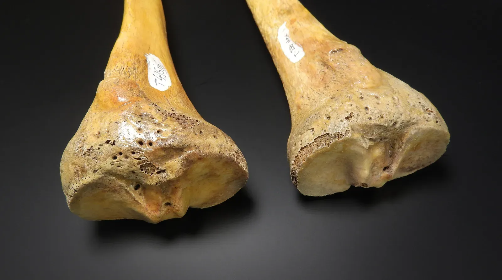
While the femur flattens front-to-back, the shin bone tells a mirror-image story. Long-distance walkers frequently develop platycnemia – a side-to-side flattening of the tibial shaft that gives the bone an almost blade-like cross-section instead of the typical triangular shape.
This adaptation shows up disproportionately in populations known from historical and ethnographic records to have covered vast territories on foot: nomadic herders, hunter-gatherer foragers, and pre-industrial messengers. The tibia essentially reshapes itself to resist the specific twisting loads produced by uneven ground, hills, and sustained forward propulsion.
What makes this sign so useful forensically is its specificity. Platycnemia doesn’t appear from sitting, standing still for long shifts, or even moderate daily activity – it requires sustained, repetitive loading over years, sometimes decades. Bioarchaeologists treat a genuinely platycnemic tibia paired with a robust, platymeric femur as one of the strongest combined indicators of a high-mobility lifestyle in the entire skeleton.
#4 – The Fibula Reveals What the Other Leg Bones Miss
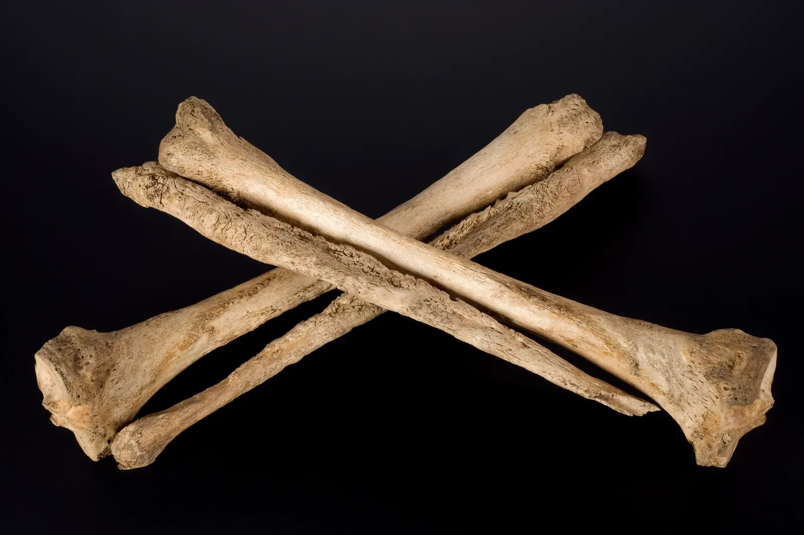
The fibula, the thin secondary bone running alongside the shin, gets almost no attention in casual anatomy conversations. It doesn’t bear much body weight, so it’s easy to assume it stays quiet no matter what a person’s life looked like. Researchers studying mobility patterns increasingly disagree, arguing it’s an overlooked mobility indicator hiding in plain sight.
A dedicated body of research now exists specifically because standard measurements were missing something. Academic literature on ancient locomotion includes entire chapters investigating lower limb structural rigidity through the cross-sectional properties of the fibular midshaft, in samples spanning the Late Upper Paleolithic through the Medieval period – specifically flagging fibular robusticity as an under-considered piece of the puzzle.
The fibula anchors major calf muscles responsible for propulsion and stabilization on uneven terrain, meaning a thickened, reinforced fibula often shows up in skeletons that logged serious mileage across rough or hilly landscapes rather than flat, easy routes. Most forensic assessments jump straight to the tibia and femur – but skipping the fibula, many specialists now argue, means missing real evidence sitting right next to it.
#5 – Muscle Attachment Sites Are Dramatically Enlarged
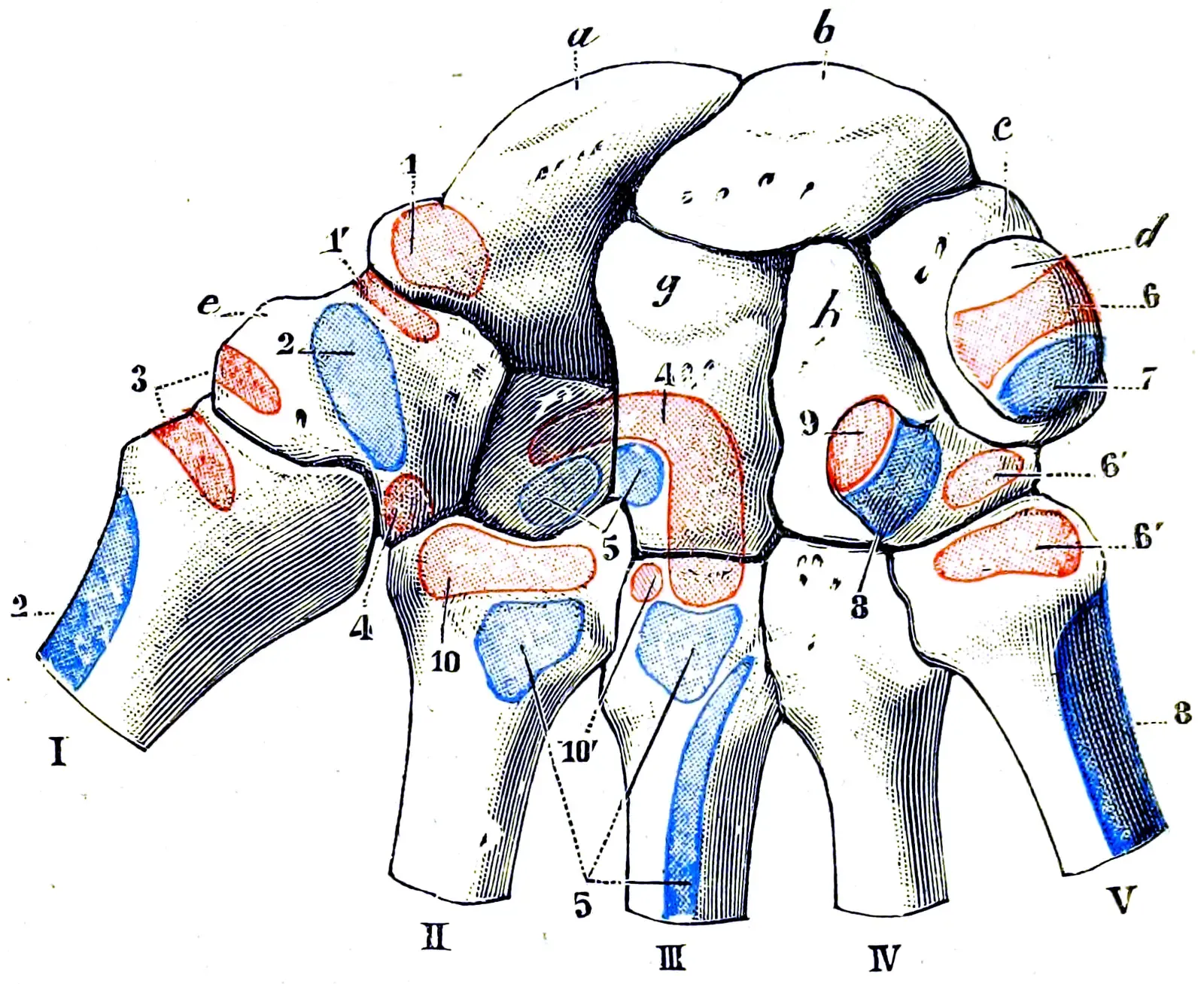
Every muscle in the human body attaches to bone at a specific point called an enthesis, and repeated, forceful contraction leaves a permanent mark there. In long-distance walkers, these marks cluster overwhelmingly in the lower limb – around the hip, knee, and calf.
Researchers scoring skeletal remains with the Coimbra method have found that individuals with heavier cumulative mechanical loading, such as modern endurance athletes, show significantly larger entheseal areas at the gluteus medius, gluteus maximus, vasti, gastrocnemius, and soleus than sedentary comparison groups, across nearly every muscle measured.
Translation: the muscles responsible for hip stabilization, knee extension, and propulsive push-off leave craters and ridges on bone when they’ve been worked relentlessly for years. A skeleton showing enlarged, roughened attachment sites specifically at the gluteus medius and soleus paints a very specific picture – someone whose hips and calves were doing serious, repetitive work day after day, exactly what sustained walking demands.
#6 – Osteoarthritis Clusters in the Hip, Knee, and Ankle
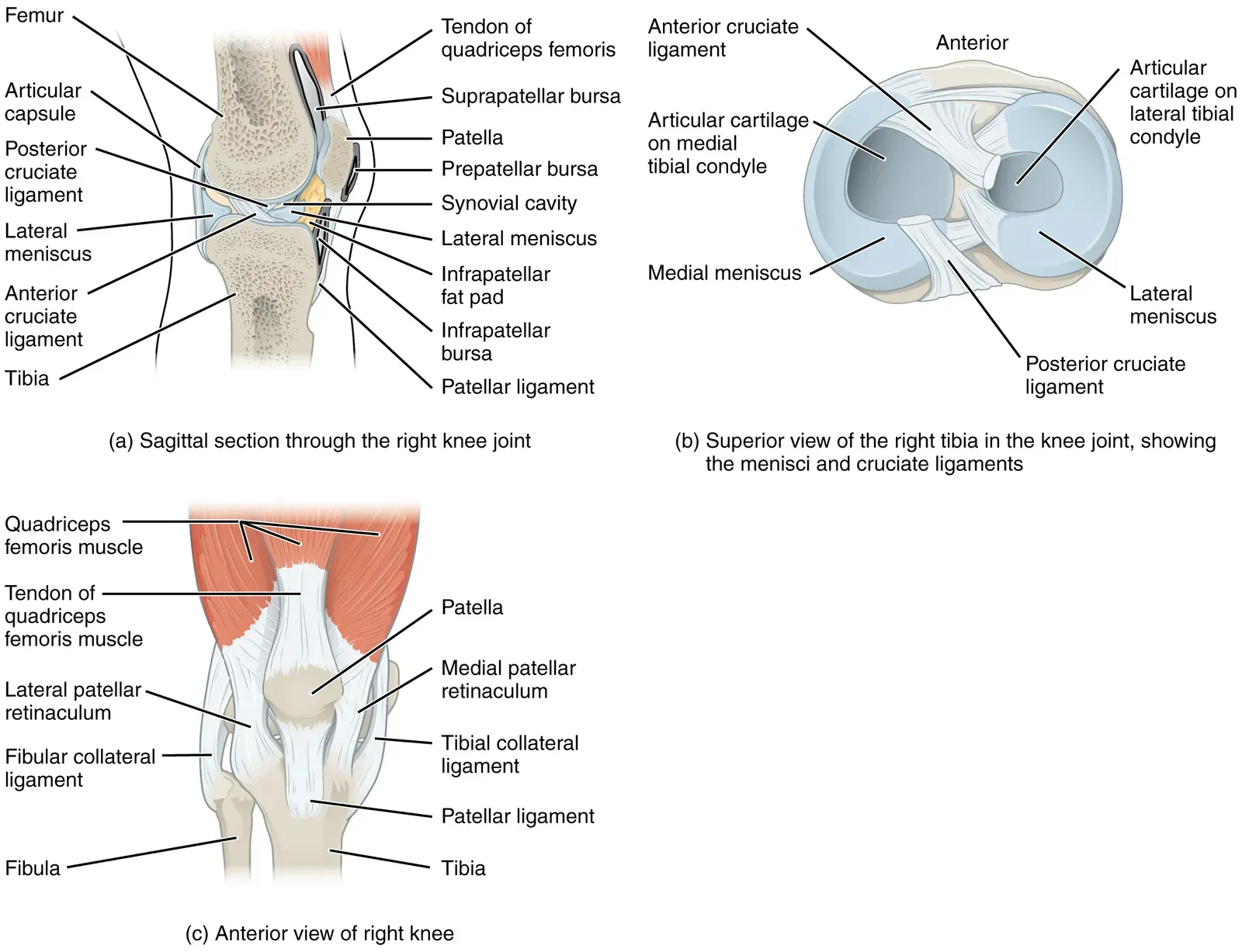
Osteoarthritis shows up in nearly every aging skeleton to some degree, so on its own it’s not a smoking gun. But the pattern matters enormously. When arthritis clusters specifically and severely in the hip, knee, and ankle – while the wrists, elbows, and shoulders stay comparatively pristine – that’s a very different story than generalized wear from age.
This localized pattern reflects decades of repetitive compressive loading concentrated in the joints that bear the brunt of a walking gait. The cartilage in these three joints wears down faster than biological aging alone would predict, leaving bone-on-bone polishing, joint margin lipping, and eburnation – a glassy bone-on-bone shine – visible even centuries later.
Forensic anthropologists specifically check whether the arthritic severity is symmetrical across both legs or lopsided. Symmetrical, severe lower-limb arthritis paired with comparatively healthy upper-limb joints is a textbook combination pointing toward a life spent moving on foot rather than performing seated or upper-body labor.
#7 – Healed Stress Fractures Riddle the Feet
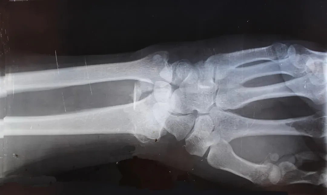
Military medicine actually coined a nickname for this exact injury: the “march fracture.” It refers to a hairline stress fracture, most commonly in the second or third metatarsal of the foot, caused by repetitive impact rather than a single traumatic event.
In skeletal remains, these fractures show up as small, healed calluses of bone – evidence the body repaired damage that built up gradually rather than all at once. A skeleton riddled with multiple healed metatarsal stress fractures is essentially carrying a written record of thousands of miles logged on hard or uneven ground.
What makes this sign compelling is timing. Stress fractures don’t happen from occasional walks; they require sustained, repetitive loading beyond what bone can immediately adapt to, followed by partial healing, followed by re-injury as the walking continued. Finding several healed fractures at different stages in the same foot suggests a chronic, ongoing pattern rather than one unlucky incident.
Worth Knowing
- Military physicians first described march fractures in soldiers logging long distances on foot.
- The second and third metatarsals absorb the most repetitive impact during a walking stride.
- These fractures form gradually, unlike a single traumatic break.
- Multiple healed fractures at different stages in one foot point to a chronic, ongoing walking pattern.
#8 – Schmorl’s Nodes Reveal a Life of Carrying, Not Just Walking
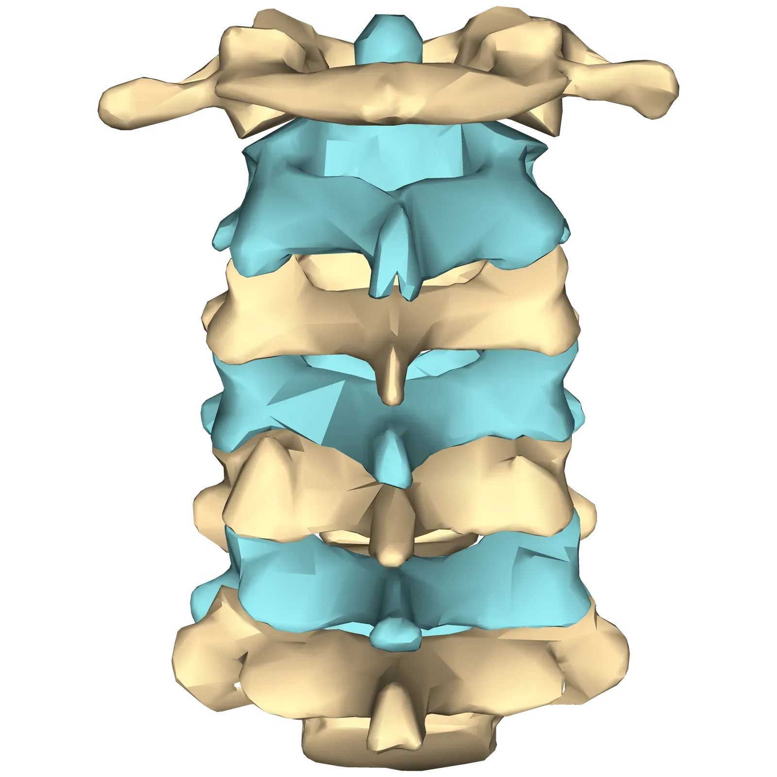
Long-distance walking rarely happens with empty hands. Historically, it meant carrying water, tools, goods, food, or children across those same enormous distances, and the spine absorbs that compounded load with every single step.
Schmorl’s nodes are small depressions that form when the soft disc material between vertebrae herniates into the bone of the vertebral body itself, usually from repeated compressive stress. Skeletons that combine heavy Schmorl’s node clustering in the lower back with clear lower-limb mobility markers suggest someone who wasn’t just walking – they were walking while loaded down.
Bioarchaeological research on horse-riding populations found a similarly instructive pattern: entheseal changes at the coxal bone, femur, tibia, and calcaneus, alongside intervertebral disc herniations at the thoracolumbar junction and ovalization of the hip socket. Walkers carrying loads over distance show a parallel spinal signature, concentrated exactly where the trunk absorbs the jolting, repetitive shock of a loaded gait.
#9 – The Hip Socket Itself Warps Out of Round
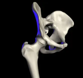
The acetabulum, the cup-shaped socket where the femur meets the pelvis, is supposed to stay a fairly consistent round shape throughout adulthood. In skeletons with a lifetime of extreme mobility, it doesn’t always stay that way.
Researchers studying populations with intensely repetitive lower-limb activity have documented actual ovalization of the acetabulum – a subtle but measurable shift away from a perfectly circular socket, caused by years of repetitive, directionally-biased loading on the hip joint. This often shows up alongside matching remodeling at the femoral neck in the same individuals.
This is one of those signs that sounds subtle in a textbook but is actually quite striking to trained eyes. A perfectly round hip socket suggests varied, balanced activity. An oval, remodeled one suggests a joint that spent years absorbing force from essentially the same repetitive motion, over and over, for a very long time – precisely the biomechanical fingerprint of habitual, sustained walking.
#10 – The Sacroiliac Joint Shows Wear It Was Never Built For
Tucked at the back of the pelvis, where the spine meets the hip bones, the sacroiliac joint doesn’t move much in most people. It’s built more for stability than mobility. But in skeletons belonging to people who covered enormous distances on foot, this joint sometimes shows wear patterns that shouldn’t exist in a “normal” aging profile.
Most people don’t realize the sacroiliac joint is essentially a shock absorber for the entire trunk during walking, transferring force between the legs and spine with every single stride. Chronic, high-volume walking over uneven or hilly terrain places repetitive stress on this joint that ordinary daily living simply doesn’t replicate.
Combined with the acetabular changes covered earlier, unusual sacroiliac wear helps anthropologists distinguish a genuine lifetime walker from someone who was simply arthritic from general aging. It’s rarely used alone as evidence – but stacked alongside femoral robusticity, tibial flattening, and hip joint changes, it becomes a meaningful piece of a much larger mobility puzzle.
#11 – The Leg Bones Grow Denser Exactly Where It Counts
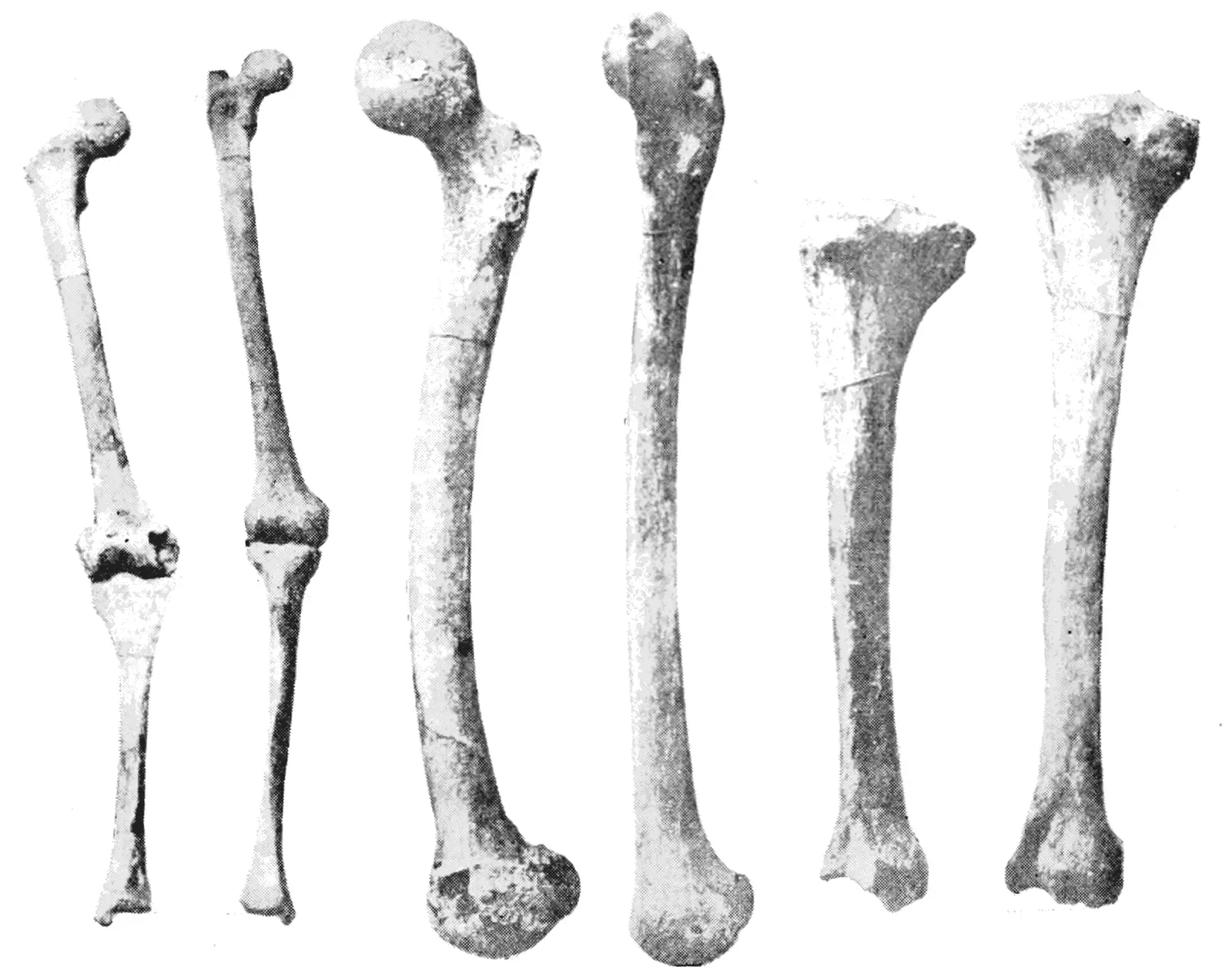
Wolff’s Law, a foundational principle in skeletal biology, states that bone remodels itself in direct response to the mechanical loads placed on it. Walk relentlessly for decades, and the leg bones respond by laying down denser, thicker cortical bone exactly where the loading is greatest.
This isn’t the same as simply having strong bones from genetics. It’s measurable, load-specific densification that shows up disproportionately in the femur and tibia rather than throughout the entire skeleton. Researchers comparing populations with different subsistence and mobility strategies have consistently found that this density signature tracks with lifestyle far more closely than it tracks with ancestry.
Interestingly, the relationship isn’t always as simple as “more walking equals denser bone” in every context. Comparisons between preagricultural and agricultural populations suggest sedentary societies can actually show greater size and rigidity in the long bones than highly mobile ones, possibly because agricultural life involves a wider variety of movement types overall. That surprises a lot of people – and it’s exactly why anthropologists never rely on bone density alone. It always gets cross-checked against the other signs on this list.
Quick Compare
- High-mobility foragers: dense, load-specific cortical bone concentrated in the femur and tibia.
- Sedentary agricultural populations: sometimes show greater overall long-bone rigidity, likely from varied movement types.
- Takeaway: bone density alone can mislead, which is why it’s always cross-checked against the other signs on this list.
#12 – One Leg Is Noticeably Stronger Than the Other
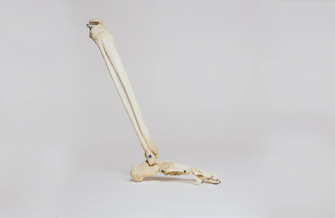
A skeleton that walked enormous, repetitive distances doesn’t always show perfectly matched legs. In populations that regularly carried loads on one side, worked consistently on sloped or cambered paths, or favored a dominant leg for balance on uneven ground, measurable asymmetry shows up between the left and right femurs and tibiae.
This asymmetry gets measured the same way overall robusticity does – through cross-sectional geometry comparing bending strength side to side. A meaningful strength difference between otherwise matched limb bones is a red flag that something habitual and repetitive, rather than random, shaped that person’s gait.
This is also one of the more debated signs among specialists, and deliberately so. Some researchers argue pronounced asymmetry says more about occupation-specific tasks – carrying water on one hip, herding on one side, favoring a walking stick – than about total distance walked. Others maintain that any sustained, high-mileage lifestyle on natural terrain almost always produces some degree of asymmetry, simply because no landscape is perfectly even underfoot. Either way, it’s a sign specialists never ignore.
#13 – The Whole Skeleton Matches a Known Walking Population
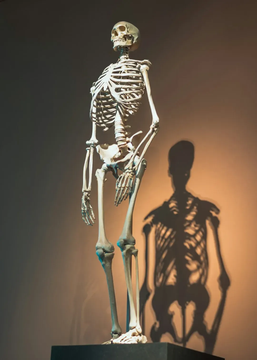
The most convincing sign isn’t any single bone – it’s the whole-body pattern matching populations bioarchaeologists already know walked constantly. Comparative studies of hunter-gatherer and forager populations, including detailed work on Middle Holocene foragers of Siberia’s Cis-Baikal region, have built a reference library of exactly what a lifetime of habitual, high-volume mobility looks like across an entire skeleton, not just one bone.
Turns out, the real diagnostic power comes from stacking every sign covered in this article together: robust, flattened femurs; platycnemic tibiae; a reinforced fibula; enlarged entheses at the hip and calf; joint-specific arthritis; healed stress fractures; spinal compression damage; hip and sacroiliac remodeling; targeted bone density; and leg asymmetry, all appearing in the same individual.
No single marker proves a person walked extraordinary distances throughout their life. But when a skeleton checks off eight, nine, or ten of these thirteen signs simultaneously, bioarchaeologists consider that about as close to a signed confession as bone can offer. This is precisely how researchers have confidently identified nomadic herders, long-distance foragers, and pre-modern messengers and traders from skeletal remains alone, sometimes thousands of years after the fact.
At a Glance
- Robust, flattened femur paired with a platycnemic tibia
- Reinforced fibula with a thickened midshaft
- Enlarged entheses at hip and calf muscle attachments
- Joint-specific arthritis in the hip, knee, and ankle
- Healed stress fractures plus spinal or hip-socket remodeling
The Bottom Line
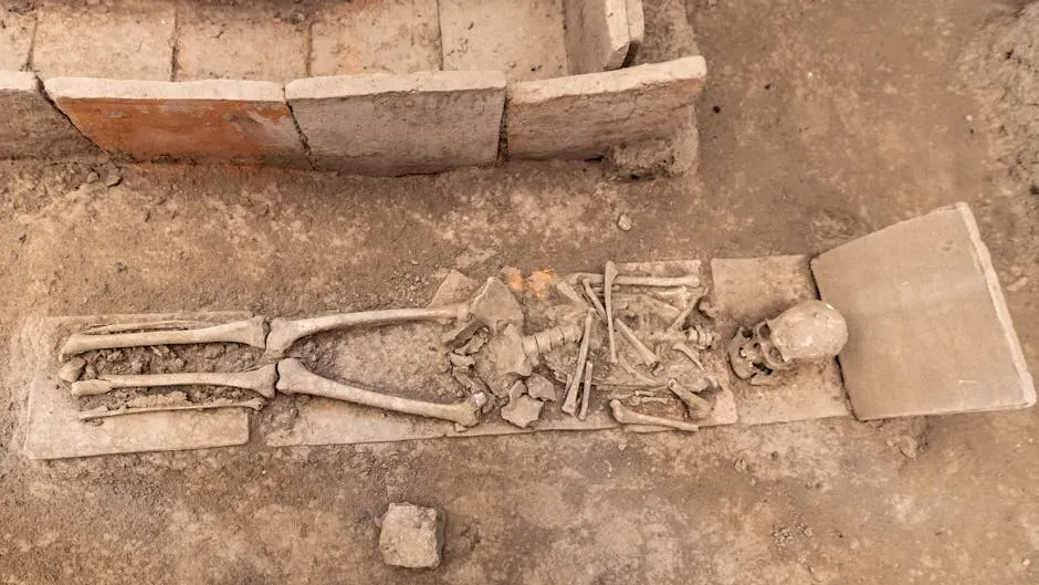
Bone doesn’t lie, and it doesn’t forget. A lifetime of walking enormous distances leaves a permanent, layered signature across the femur, tibia, fibula, hips, spine, and feet that no amount of time erases. Individually, these thirteen signs can be ambiguous. Stacked together, they become one of the most reliable behavioral fingerprints in all of skeletal science.
What strikes us most is how consistently the evidence contradicts the lazy assumption that “big, strong bones” simply mean a hard laborer. The real story is far more specific, and far more human – it’s written in flattened shins, oval hip sockets, and healed fractures that took years to accumulate. Which of these thirteen signs surprised you the most? Drop it in the comments.