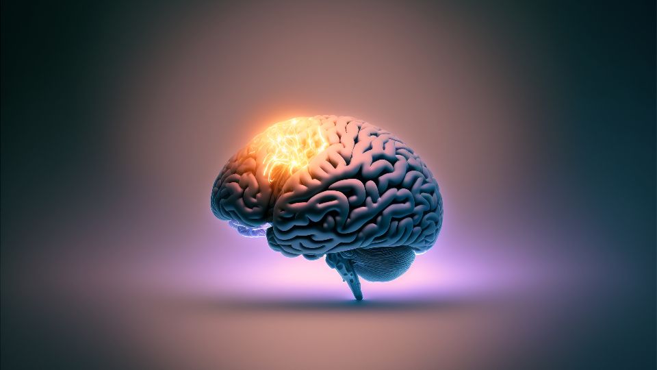
When you imagine death, you probably picture a clear on–off switch: the heart stops, the monitor flatlines, and everything in the brain goes dark at the same moment. That picture feels intuitive, but biology does not respect clean lines or cinematic timing. Instead, your body unravels in stages, and your brain is one of the last, most stubborn parts of you to let go.
In the last decade, scientists have used more sensitive tools and bold experiments to show that certain forms of brain activity can persist for hours after what doctors call clinical death. That does not mean consciousness survives or that resurrection is around the corner; it means your brain is a complex ecosystem of cells and chemicals that wind down in slow motion, not all at once. When you look closely, you see a hidden twilight period where neurons struggle, glial cells hustle, and energy systems flicker in and out before finally failing.
The Moment of Clinical Death Is Not the Moment Your Brain “Dies”

When a doctor pronounces clinical death, they are usually marking the moment your heart has stopped beating and blood is no longer circulating oxygen through your body. For practical and legal purposes, this is the line that matters in an emergency room. But at the cellular level, especially in your brain, things are nowhere near that simple. Your neurons do not instantly vanish; they begin a desperate fight for survival that can last minutes to hours, depending on conditions.
You can think of it like pulling the plug on a huge, busy city at night. The power grid might go down, but car headlights, backup generators, and battery-powered devices keep flickering for a while. In your brain, the power grid is the continuous flow of oxygenated blood, and when it fails, different brain regions shut down at different speeds. Areas that are starved of oxygen and energy fail quickly, while others with different metabolic demands or better local reserves keep limited activity going for surprisingly long stretches of time.
How Neurons Lose Consciousness Long Before They Lose All Activity
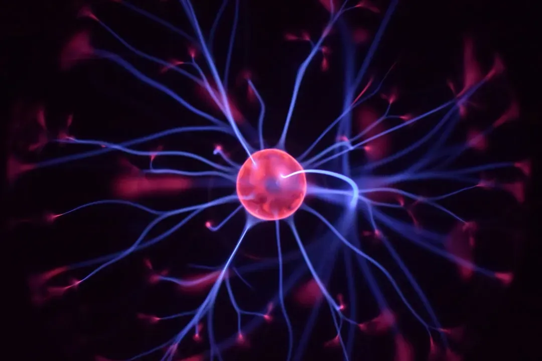
Consciousness is fragile. You lose it under anesthesia, during deep sleep, or when blood flow briefly drops. After your heart stops, global networks that support awareness collapse within seconds to a few minutes. The synchronized electrical patterns you see on an electroencephalogram, the ones associated with being awake and responsive, quickly break apart. From your point of view, if you could experience it, there is no long drawn-out awareness; the lights go out fast.
But just because consciousness has vanished does not mean every neuron is silent. Individual cells can keep firing, small clusters can show local activity, and certain pathways might still conduct impulses for a time. This is like a stadium after a game: the big show is over, the crowd is gone, but you still have cleaners moving around, lights in side rooms, and a few staff members talking. When researchers look with sensitive electrodes or molecular tools, they can detect these smaller, scattered signals that persist long after any meaningful awareness is possible.
The Slow Collapse of Energy: Oxygen, ATP, and Ion Pumps
Your brain is an energy addict. Even though it makes up only a small fraction of your body weight, it uses a surprisingly large share of your available energy at rest. That energy is mostly spent on running tiny molecular pumps in your neurons that maintain differences in ion concentrations across their membranes. Those differences are what allow electrical signals – action potentials – to exist at all. When your heart stops, oxygen delivery falls to zero, and your brain’s energy factories, the mitochondria, can no longer keep up.
At first, your cells rely on the last bits of stored fuel and oxygen. Adenosine triphosphate, the main energy currency, starts to drop, but not instantly. As ATP dwindles, those ion pumps begin to fail, and neurons can no longer hold their carefully balanced gradients. You can picture a dam that once kept water neatly separated; as the power for the floodgates disappears, everything leaks and mixes. This progressive breakdown leads to electrical silence on a large scale, yet some pockets of cells, especially those with slightly better reserves or different metabolic needs, may limp along, producing weak or sporadic electrical signals for quite some time.
Glial Cells: The Unsung Players Still Working After the Heart Stops
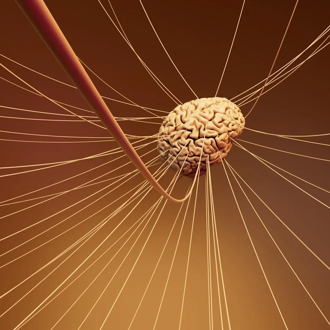
When you think about brain activity, you probably picture neurons, but they are only one part of the story. Your brain is full of glial cells – astrocytes, microglia, oligodendrocytes – that support, protect, and modulate neural function. Unlike neurons, glial cells do not mainly communicate through rapid electrical spikes; they work with slower waves of chemical signals, calcium pulses, and structural changes. These processes are more resilient during the early phases after blood flow stops, so glial cells can stay active even while neurons are failing.
Astrocytes, for instance, help regulate ion balance and neurotransmitter levels, and they can respond to rising stress signals during the dying process. Microglia, the immune-like cells of your brain, may begin shifting into an activated state when they sense damage. Because these glial responses unfold over minutes to hours rather than milliseconds, you can still see measurable activity in certain brain regions, especially at the microscopic level, long after classic electrical patterns have gone flat. It is less like the frantic chatter of a live concert and more like a slow, tense negotiation in the back rooms as the building shuts down.
Ionic Waves, Spreading Depolarization, and the Final Electrical Storm
As your brain’s ion pumps fail, something dramatic happens: large regions of cortex can experience what researchers call spreading depolarization or a terminal wave. During this event, the controlled separation of ions across neuronal membranes collapses, and a wave of electrical and chemical change sweeps across the tissue. This is not organized, information-rich activity; it is more like a massive short-circuit that marks the point of no return for those cells if blood flow is not restored.
What makes this fascinating is that under certain conditions, such waves do not occur immediately when blood flow stops. There can be a delay of several minutes, sometimes longer, during which the tissue sits in a precarious state. Even after the big wave passes, some residual ionic movements, molecular signaling, and glial responses can continue locally for a while. If you are monitoring closely enough, especially deep in the tissue rather than at the scalp surface, you may still detect these shifts and interpret them as remaining activity, even though the neurons have lost any realistic chance of recovery.
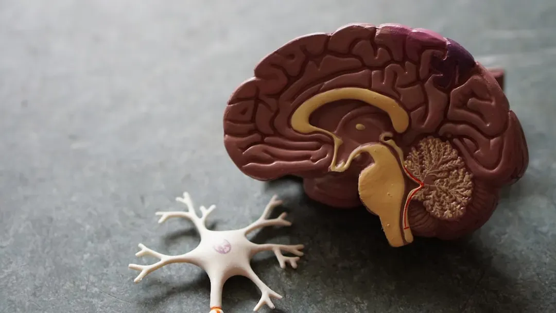
Why Brain Tissue Can Sometimes Be “Revived” Hours After Death in the Lab
In experimental settings, scientists have taken brain tissue from animals after death and perfused it with oxygenated solutions to study how cells respond. In some high-profile work, researchers were able to restore certain cellular functions and synaptic responses hours after circulation had stopped, although they did not restore consciousness or normal brain rhythms. For you, the key lesson is that neurons and glia can be more resilient than you might assume, especially when cooled or handled in ways that slow down the damage.
Think of it like finding a plant that looks wilted and lifeless after a long dry spell. If you water it quickly enough and the roots are not completely destroyed, some leaves may perk up. In a similar way, brain cells can still hold partial structure and potential even when the organism as a whole is long past any chance of survival. Under carefully controlled lab conditions, scientists can coax some of that hidden capacity back into action. That lingering potential is part of why you see measurable activity in parts of the brain hours after clinical death in research settings, even though it does not translate into any meaningful recovery for a person.
What This Ongoing Activity Means for Organ Donation and Resuscitation
Because your brain does not die all at once, medicine has had to draw practical lines for decisions about organ donation and attempts at resuscitation. Heart-beating donors, for example, are usually declared dead based on neurological criteria that reflect irreversible loss of the capacity for consciousness and breathing, even if some cells in the brain are technically still alive or metabolically active. When you learn that some brain activity can linger, it might sound unsettling, but the key is to understand that not all activity is equal.
The kind of scattered, cellular-level processes that persist after clinical death do not support awareness, memory, or anything you would recognize as selfhood. Instead, they are more like the last flickers in a complex machine winding down. At the same time, knowing that some brain regions remain responsive for a period opens doors for better resuscitation strategies and timing decisions. Cooling, oxygenation, and careful management of blood flow can potentially extend the window during which brain cells can be saved, which matters a lot if you ever face a cardiac arrest where minutes make the difference between recovery and devastating injury.
Separating Science From Myth: No, This Does Not Prove Consciousness Survives Death
When you hear that parts of the brain remain active for hours after clinical death, it is easy to jump to big philosophical or spiritual conclusions. You might wonder if consciousness lingers, if people are aware of what happens around them, or if this is evidence that your mind can float free from your body. The science you have so far does not support those stories. What persists is cellular metabolism, stress responses, and disorganized electrical and chemical activity, not the coordinated patterns associated with thinking or awareness.
In other words, the fact that individual circuits twitch after the system has crashed is not proof that the system is still running. You can unplug a computer and still find residual charges on chips for a while, but the operating system is gone. Recognizing this helps you keep your expectations grounded: the brain’s twilight phase is biologically rich and scientifically important, but it is not an extension of lived experience. Accepting that difference can be oddly comforting, because it lets you appreciate the complexity of your biology without blurring the line between the end of consciousness and the slower end of cells.
Conclusion: A Brain That Dies in Stages, Not in an Instant
When you pull together all these threads, you see death differently. Your brain is not a lightbulb that switches off the instant your heart stops; it is more like a vast city losing power neighborhood by neighborhood, with emergency crews still moving in the dark, systems failing at different speeds, and some small pockets of function hanging on far longer than you would expect. Clinical death marks a clear medical and legal boundary, but beneath that label lies a long, intricate biological unwinding filled with delayed waves, glial labor, and last-ditch cellular struggles.
For you, understanding this slow fade does two things. It keeps your thinking grounded and protects you from over-interpreting the lingering sparks of activity as signs of hidden awareness, while also deepening your respect for how tenacious and elaborate your brain really is. That knowledge shapes conversations about resuscitation, organ donation, and what it means to die with dignity in a world where technology can sometimes nudge the boundaries. As you sit with the idea of a brain that dies in stages, not in an instant, you might quietly ask yourself: does this make death feel stranger, or does it somehow make it a little easier to understand?