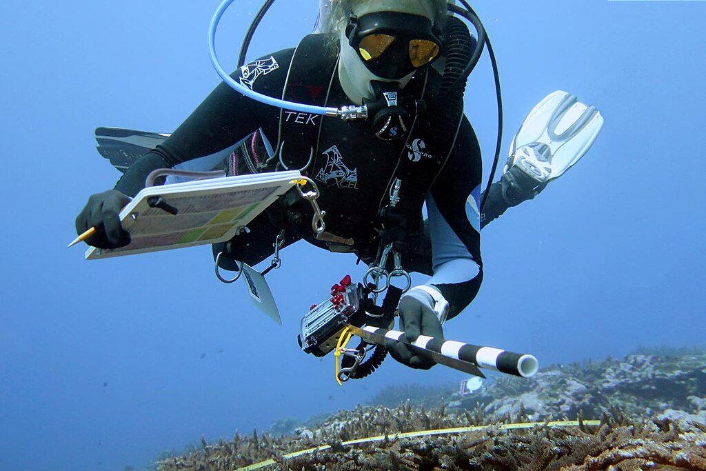
A new diver-operated microscope is revolutionizing coral research by allowing scientists to observe photosynthesis and microalgae behavior directly in the ocean. Developed by UC San Diego’s Scripps Institution of Oceanography, the Benthic Underwater Microscope Imaging PAM (BUMP) offers unprecedented access to coral bleaching and how they respond to environmental stress.
A Technological Leap for Coral Science
The BUMP system uses pulse amplitude modulated (PAM) light to measure photosynthetic efficiency within coral tissues. It captures high-resolution images and fluorescence data without disturbing the reef, enabling scientists to visualize how individual microalgae cells function inside coral polyps. This non-invasive approach marks a major advance in field-based coral health assessment.
Seeing Coral Behavior Up Close
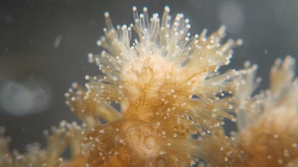
During field tests in Hawaii, the Red Sea, and Palmyra Atoll, researchers observed dynamic coral behaviors, including tentacle contractions and particle capture, that were previously undocumented. The microscope revealed red fluorescence from chlorophyll in macroalgae and cyan/green fluorescence from coral proteins, offering clues about coral metabolism and stress responses.
Implications for Conservation
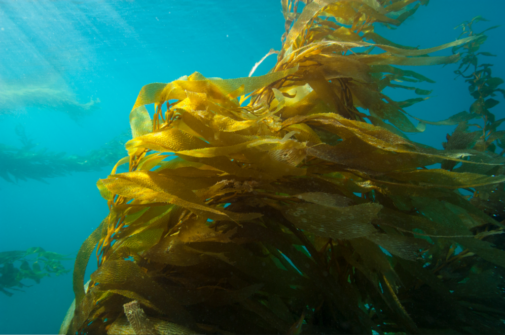
By detecting early signs of bleaching and mapping photosynthesis in situ, the BUMP tool could help scientists predict coral decline before irreversible damage occurs. Its portability and precision make it ideal for monitoring reefs in remote locations, and its applications extend to other photosynthetic marine organisms like juvenile kelp.
Conclusion
The BUMP microscope brings the lab to the reef, illuminating coral biology at scales never before seen. As marine ecosystems face mounting threats, this tool offers a powerful way to track coral health, decode bleaching mechanisms, and guide conservation strategies, one polyp at a time.
Source: